Acute appendicitis is one of the most common surgical emergencies worldwide and remains a frequent indication for emergency abdominal surgery. Although advances in clinical assessment, laboratory investigations, imaging modalities, and diagnostic algorithms have improved diagnostic accuracy, distinguishing appendicitis from other causes of right lower quadrant pain remains challenging. Consequently, diagnostic laparoscopy continues to play an important role in the evaluation and management of patients with suspected appendicitis, particularly when the diagnosis remains uncertain.
A persistent intraoperative dilemma arises when the appendix appears macroscopically normal during surgery performed for suspected appendicitis. There is currently no universal consensus on whether such an appendix should be removed or left in situ after alternative causes of the patient’s symptoms have been excluded.1 An international survey of surgeons identified the absence of clear clinical guidelines and concern about missing endoluminal or early appendicitis as the principal reasons for performing appendectomy despite a normal macroscopic appearance.2
Histopathological diagnosis of acute appendicitis has traditionally relied on the presence of transmural inflammation.3 However, some pathology departments also recognise isolated mucosal ulceration or mucosal inflammation as evidence of early appendicitis.4 Consequently, inflammation confined to the mucosa may not be apparent on macroscopic inspection of the serosal surface, raising concerns that a macroscopically normal appendix may harbour clinically significant pathology. Failure to identify and treat occult appendicitis may result in delayed diagnosis, perforation, intra-abdominal abscess, diffuse peritonitis, reoperation, prolonged hospitalisation, and increased healthcare costs.
Conversely, routine removal of all macroscopically normal appendices is not without consequences. Negative appendectomy has been associated with unnecessary exposure to anaesthesia and operative risks, postoperative morbidity, longer hospital stay, and increased healthcare expenditure compared with appendectomy performed for confirmed uncomplicated appendicitis.5 Unnecessary surgery also imposes avoidable physical, psychological, and economic burdens on patients, families, and healthcare systems.6 Therefore, the decision to remove or preserve a macroscopically normal appendix requires balancing the risk of missed disease against the harms associated with unnecessary surgery.
Current recommendations reflect this uncertainty. The Society of American Gastrointestinal and Endoscopic Surgeons (SAGES) advises that, after excluding other causes of abdominal pain, removal of a macroscopically normal appendix may be considered on an individual patient basis.7 Supporting this cautious approach, Lee et al. reported that 13.3% of patients in whom a macroscopically normal appendix was left in situ subsequently required reoperation, of whom 4.17% had histologically confirmed appendicitis.8 However, the available evidence remains inconsistent and is largely derived from small observational studies with variable definitions of both a macroscopically normal appendix and histological appendicitis.
Although several studies have examined the relationship between intraoperative macroscopic assessment and subsequent histopathological findings, the reported incidence of occult appendicitis varies considerably, and the overall burden of clinically significant pathology remains uncertain. A comprehensive synthesis of the available evidence is therefore needed to inform surgical decision-making, guideline development, and future research.
Accordingly, this systematic review and meta-analysis aimed to determine the pooled incidence of histologically confirmed appendicitis and other clinically significant pathology in macroscopically normal appendices removed during surgery for suspected appendicitis. By quantifying the diagnostic limitations of macroscopic assessment, this review seeks to provide evidence to support clinical decision-making and inform future health policy and research evaluating the balance between unnecessary appendectomy and missed appendiceal disease.
Methods
Protocol registration and reporting
This systematic review and meta-analysis was prospectively registered in the International Prospective Register of Systematic Reviews (PROSPERO; CRD42023397449) and conducted in accordance with the Preferred Reporting Items for Systematic Reviews and Meta-Analyses (PRISMA 2020) guidelines.9
Search strategy
A comprehensive literature search was undertaken in PubMed, MEDLINE, Scopus, the Cochrane Central Register of Controlled Trials (CENTRAL), the Cochrane Library, the World Health Organization International Clinical Trials Registry Platform (WHO ICTRP), and Google Scholar to identify relevant studies published up to 1 March 2023.
The search strategy incorporated combinations of the following keywords and Medical Subject Headings (MeSH), where applicable: “macroscopically normal appendix”, “negative appendectomy”, “false negative appendectomy”, “appendicitis”, “diagnostic laparoscopy”, “laparoscopy”, and “appendectomy”. Reference lists of all eligible studies were manually screened to identify additional relevant publications (Appendix 1, Online Supplementary Document)
Eligibility criteria
Published full-text experimental and observational studies evaluating histopathological findings in macroscopically normal appendices removed during surgery for suspected appendicitis were eligible for inclusion. No language restrictions were applied.
Review articles, editorials, letters, conference abstracts, case reports, animal studies, and studies for which the full text could not be retrieved were excluded.
Study selection
All retrieved records were imported into Covidence software for study management and duplicate removal.
Two reviewers (R.Sh and H.M) independently screened titles and abstracts for eligibility. Potentially relevant studies subsequently underwent full-text assessment by the same reviewers. Disagreements were resolved through discussion, and where consensus could not be reached, a third reviewer (A.A) acted as an adjudicator.
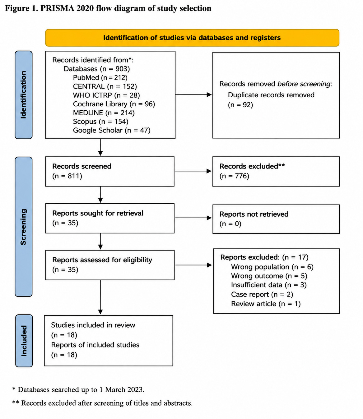
The study selection process is summarised in the PRISMA flow diagram (Figure 1).
Data extraction
Data extraction was independently performed by R.Sh and verified by H.M and A.A using a standardized data extraction form.
The following information was extracted from each study:
-
year of publication;
-
study design;
-
study objectives;
-
sample size;
-
participant characteristics (including age and sex);
-
intraoperative findings;
-
management of the macroscopically normal appendix;
-
histopathological findings; and
-
incidence of appendicitis or other significant pathology in macroscopically normal appendices.
Quality assessment
Methodological quality and risk of bias were independently assessed by two reviewers (R.Sh and H.M), with disagreements resolved by discussion or, when necessary, consultation with a third reviewer (A.A).
Observational studies were assessed using the Modified Newcastle-Ottawa Scale (NOS), a validated tool for evaluating the methodological quality of non-randomized studies (Appendix 2, Online Supplementary Document).10 The assessment considered three domains: selection of study groups, comparability between groups, and ascertainment of the exposure or outcome of interest.
Statistical analysis
The primary outcome was the pooled incidence of histologically confirmed acute appendicitis in macroscopically normal appendices.
A secondary analysis estimated the pooled incidence of overall clinically significant pathology identified on histopathological examination despite a macroscopically normal intraoperative appearance.
Pooled incidence estimates were calculated using random-effects meta-analysis and are presented as proportions with corresponding 95% confidence intervals (CIs). Given the anticipated clinical and methodological heterogeneity across the included studies, the DerSimonian-Laird random-effects model was used to estimate between-study variance.
Statistical heterogeneity was quantified using the I² statistic, with values of <25%, 25%–50%, and >50% representing low, moderate, and high heterogeneity, respectively.
Publication bias was assessed through visual inspection of funnel plots and Egger’s regression test for small-study effects.
All statistical analyses were performed using Stata version 18.0 (StataCorp LLC, College Station, TX, USA).
Results
Study selection
The database search identified 903 records, of which 92 duplicate records were removed. Following title and abstract screening, 776 studies were excluded, leaving 35 articles for full-text assessment. Seventeen studies were subsequently excluded for not meeting the eligibility criteria, resulting in 18 studies being included in the final qualitative and quantitative synthesis (Figure 1).
Study characteristics
The included studies were published between 1988 and 2021 and comprised two prospective cohort studies,11,12 one retrospective cross-sectional study,13 and fifteen retrospective cohort studies. Across all studies, 1,780 patients with a macroscopically normal appendix identified during diagnostic laparoscopy or open surgery for suspected appendicitis underwent appendectomy. Histopathological findings were compared with the intraoperative macroscopic assessment in all included studies. The characteristics of the included studies are summarised in Table 2.
Quality assessment
The methodological quality of the included studies was assessed using the Modified Newcastle-Ottawa Scale (Table 1). Overall, the studies demonstrated acceptable methodological quality; however, most were retrospective observational studies and therefore remained susceptible to inherent risks of selection bias, information bias, and residual confounding.
Histopathological findings in macroscopically normal appendices
Six studies reported the presence of alternative intraoperative pathology that could potentially explain the patient’s symptoms despite the appendix appearing macroscopically normal.13,14,20–22,27 Nevertheless, appendectomy was performed in these patients. Only three of these studies analysed patients with concomitant pathology separately when estimating the incidence of appendicitis.13,20,27
Histopathological examination demonstrated substantial discordance between macroscopic appearance and microscopic findings. Acute appendicitis was the predominant abnormal histological diagnosis, accounting for 96.5% of all false-negative macroscopic assessments (Table 3). Other clinically significant pathological findings included nine neuroendocrine tumours (1.7% of all false-negative cases),11,19,20,24 one appendiceal cystadenoma,16 and one case of isolated mucosal ulceration without transmural inflammation.11 Two studies reported additional histopathological abnormalities without specifying their nature or clinical significance.13,23
Meta-analysis of histologically confirmed acute appendicitis
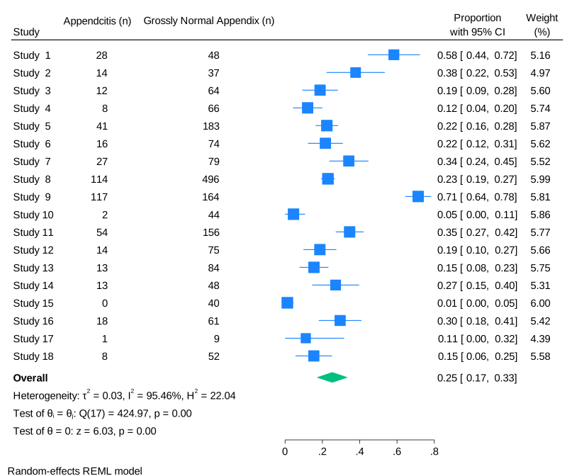
Across the included studies, the pooled incidence of histologically confirmed acute appendicitis in macroscopically normal appendices was 25% (95% CI 0.15–0.33) (Figure 2).
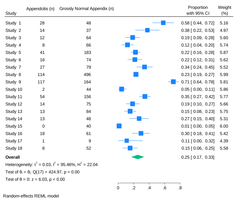
Considerable between-study heterogeneity was observed (I² = 95%), indicating substantial variability in study populations, operative assessment, histopathological definitions, and study methodology.
Meta-analysis of overall clinically significant pathology
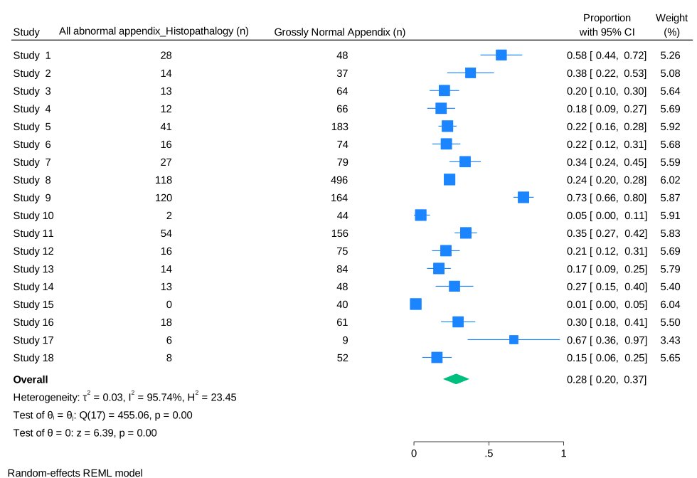
When all clinically significant histopathological abnormalities were considered, including appendicitis and other important pathological diagnoses, the pooled incidence increased to 28% (95% CI 0.20–0.37) (Figure 3).
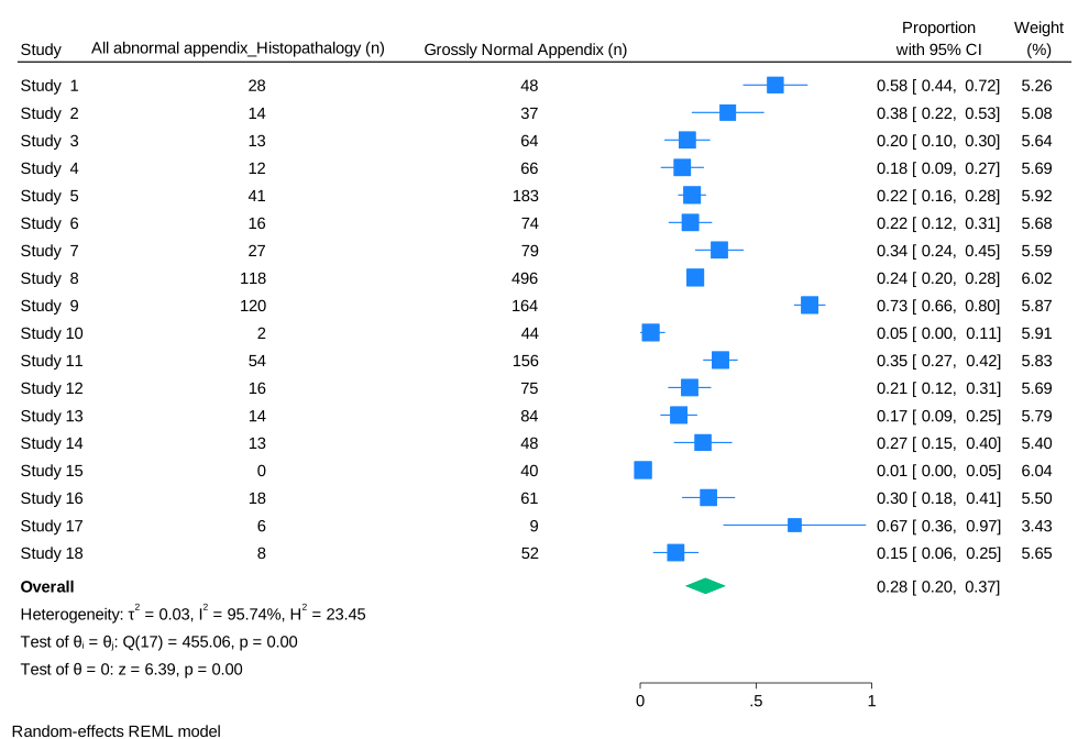
Individual study estimates varied considerably. Tartaglia et al.20 reported the highest proportion of false-negative macroscopic assessments, with 73% (95% CI 0.66–0.80) of macroscopically normal appendices demonstrating significant histopathological abnormalities. Zarandi et al.13 reported an overall false-negative rate of 67% (95% CI 0.36–0.97), although only 11% (95% CI 0.00–0.32) represented histologically confirmed acute appendicitis. Chiarugi et al.14 reported that 58% (95% CI 0.44–0.72) of macroscopically normal appendices demonstrated histopathological evidence of inflammation.
Publication bias
Visual inspection of the funnel plots did not suggest substantial publication bias. Egger’s regression test demonstrated no evidence of small-study effects (β = 1.81, standard error = 2.082; z = 0.87; p = 0.384), indicating that publication bias was unlikely to have materially influenced the pooled estimates.
Discussion
Principal findings
This systematic review and meta-analysis synthesised the available evidence on the histopathological outcomes of macroscopically normal appendices removed during surgery for suspected appendicitis. The findings demonstrate that macroscopic intraoperative assessment alone is an unreliable predictor of underlying appendiceal pathology. Approximately one-quarter of macroscopically normal appendices (25%) demonstrated histologically confirmed acute appendicitis, while more than one-quarter (28%) contained clinically significant pathology. Although acute appendicitis accounted for most abnormal histological findings, other important diagnoses, including neuroendocrine tumours and appendiceal neoplasms, were also identified. Collectively, these findings suggest that reliance on the gross appearance of the appendix alone may result in missed pathology in a substantial proportion of patients.
Comparison with previous literature
The diagnosis of acute appendicitis remains challenging despite advances in clinical assessment, laboratory investigations, and imaging. Clinical scoring systems may assist risk stratification but have limited diagnostic accuracy when used in isolation and are often more useful for monitoring disease progression in conservatively managed patients than for making definitive surgical decisions.29 Similarly, although computed tomography has substantially improved diagnostic performance and reduced negative appendectomy rates, reported diagnostic accuracy remains imperfect and its widespread use is associated with increased healthcare costs, radiation exposure, and variable availability, particularly in emergency settings.30–33
Diagnostic laparoscopy therefore continues to play an important role in the evaluation of patients with suspected appendicitis when diagnostic uncertainty persists.34,35 However, uncertainty frequently remains when the appendix appears macroscopically normal. The pooled findings of this review demonstrate considerable discordance between intraoperative macroscopic appearance and histopathological diagnosis, suggesting that visual inspection alone cannot reliably exclude early or occult appendicitis.
The wide variation observed across individual studies likely reflects differences in patient selection, surgeon experience, operative techniques, histopathological criteria, and definitions of a macroscopically normal appendix. For example, Tartaglia et al.20 reported the highest proportion of false-negative macroscopic assessments, whereas lower incidences were reported by Zarandi et al.13 and Chiarugi et al.14 These differences emphasise the lack of standardised intraoperative criteria and underscore the need for caution when interpreting macroscopic findings in isolation.
Clinical implications
The findings of this review support an individualized approach to intraoperative decision-making rather than routine preservation or routine removal of all macroscopically normal appendices. In patients in whom no alternative explanation for right lower quadrant pain is identified, appendectomy may reduce the risk of missed appendicitis and prevent subsequent complications, including perforation, intra-abdominal abscess, peritonitis, and reoperation.
Conversely, routine appendectomy carries its own risks, including operative complications, anaesthetic exposure, postoperative morbidity, and negative appendectomy. The decision should therefore be informed by the overall clinical picture, including preoperative probability of appendicitis, intraoperative findings, patient characteristics, surgeon judgement, and the availability of postoperative observation and follow-up. Rather than relying exclusively on the macroscopic appearance of the appendix, surgeons should integrate all available clinical and operative information when deciding whether appendectomy is warranted.
Health system, economic and policy implications
The implications of these findings extend beyond individual patient management and are highly relevant to healthcare resource allocation and surgical policy. The management of a macroscopically normal appendix represents a balance between two competing healthcare risks: unnecessary appendectomy and missed appendicitis.
Routine removal of macroscopically normal appendices may increase operative time, pathology workload, hospital costs, and expose some patients to avoidable surgery. Conversely, failure to remove an appendix containing occult inflammation may result in delayed diagnosis, perforation, emergency reoperation, prolonged hospitalisation, increased healthcare expenditure, and avoidable morbidity. From a health system perspective, these downstream consequences may ultimately consume substantially greater healthcare resources than the initial appendectomy itself.
The optimal management strategy is also likely to vary according to healthcare setting. In high-income countries, widespread access to high-quality imaging, pathology services, specialist surgeons, and structured postoperative follow-up may support more selective preservation of a macroscopically normal appendix. In contrast, many low- and middle-income countries continue to face delayed presentation, limited access to advanced diagnostic imaging, shortages of specialist surgical services, and restricted opportunities for timely re-evaluation. Under these circumstances, the clinical and economic consequences of missed appendicitis may be considerably greater, potentially favouring appendectomy when no alternative pathology is identified.
These findings therefore support the development of context-specific rather than universal clinical guidelines. Future recommendations should consider not only diagnostic accuracy and surgical outcomes but also healthcare capacity, resource availability, patient access to follow-up, and the economic consequences of both overtreatment and delayed treatment. Incorporating health economic evidence into future guideline development would facilitate more efficient allocation of healthcare resources while maintaining patient safety.
Strengths and limitations
This review represents, to our knowledge, the first systematic review and meta-analysis to quantify the incidence of histologically confirmed appendicitis and other clinically significant pathology in macroscopically normal appendices removed during surgery for suspected appendicitis. A comprehensive literature search, duplicate study selection, standardized quality assessment, and quantitative synthesis strengthen the validity of the findings.
Nevertheless, several limitations should be acknowledged. First, the evidence base consisted entirely of observational studies, most of which were retrospective, limiting causal inference and increasing susceptibility to selection bias and residual confounding. Second, substantial statistical heterogeneity was observed across studies, reflecting differences in patient populations, surgical techniques, definitions of a macroscopically normal appendix, and histopathological diagnostic criteria. Third, the included studies spanned more than three decades, during which imaging modalities, operative practice, and pathological classification have evolved considerably. Finally, important patient-centred outcomes, including postoperative complications, symptom resolution, readmissions, quality of life, and long-term outcomes, were inconsistently reported, precluding quantitative synthesis.
Future research
Future research should focus on well-designed prospective multicentre studies using standardized definitions of both macroscopically normal appendix and histological appendicitis. Comparative studies evaluating appendectomy versus observation should incorporate clinically meaningful outcomes, including postoperative complications, quality of life, readmissions, patient-reported outcomes, and long-term morbidity.
Importantly, future investigations should also include formal economic evaluations, such as cost-effectiveness and cost-utility analyses, to determine whether routine or selective appendectomy represents the most efficient use of healthcare resources in different health system contexts. Such evidence would strengthen future clinical guidelines and support evidence-informed policy decisions across both high-income and resource-constrained settings.
Funding
The authors received no specific funding for this work.
Authorship contributions
All authors contributed substantially to the conception and design of the study, data acquisition, analysis and interpretation, manuscript drafting and critical revision, approved the final version of the manuscript, and agree to be accountable for all aspects of the work.
Competing interests
The authors have completed the International Committee of Medical Journal Editors (ICMJE) Uniform Disclosure Form and declare that they have no competing interests.
Correspondence to:
Raya Al Shaaibi
General Surgery Residency Program
Oman Medical Specialty Board
Muscat, Oman
Email: alshaaibi.raya@gmail.com
ORCID: https://orcid.org/0000-0002-6888-3909