Cardiovascular disease (CVD) is the leading cause of death globally, with the World Health Organization estimating that CVD caused 19.8 million deaths in 2022, representing approximately 32% of all global deaths, and that more than three-quarters of cardiovascular (CV) deaths occur in low- and middle-income countries (LMICs).1 Type 2 diabetes mellitus (T2DM) is a major contributor to this burden because CVD is a principal cause of death and disability among people living with T2DM.2 The global burden of T2DM is increasing substantially, with projections suggesting that more than 1.31 billion people may be living with diabetes by 2050.3 In Africa, the International Diabetes Federation estimated that 24 million adults were living with diabetes in 2021, projected to rise to 55 million by 2045, with 54% remaining undiagnosed.4
T2DM is associated with a spectrum of CV structural and functional abnormalities, referred to as diabetic cardiomyopathy, denoting hyperglycaemia- and insulin-resistance-related CVD, characterized in early stages by diastolic dysfunction and later by overt heart failure in the absence of other dominant causes such as coronary artery disease or hypertension.5 Conventional echocardiographic assessment relies heavily on left ventricular ejection fraction (LVEF), but ejection fraction may remain preserved despite early myocardial dysfunction. Speckle-tracking echocardiography allows assessment of myocardial deformation using global longitudinal strain (GLS), which can detect subclinical left ventricular (LV) systolic dysfunction before overt reduction in LVEF becomes apparent.6
Albuminuria, commonly measured using the urinary albumin-to-creatinine ratio (UACR), is an established marker of kidney disease and diabetic kidney involvement. The Kidney Disease: Improving Global Outcomes guideline classifies albuminuria into normal to mildly increased (A1), moderately increased (A2), and severely increased (A3) categories based on UACR thresholds.7 In diabetes, albuminuria reflects abnormalities of the glomerular filtration barrier and has been linked to glomerular endothelial injury.8 Beyond its renal significance, albuminuria is also a CV risk marker. In the Heart Outcomes Prevention Evaluation study, albuminuria was associated with increased risk of CV events, CV death, and heart failure in individuals with and without diabetes.9 A recent review further summarized albuminuria as being associated with coronary artery disease, stroke, heart failure, arrhythmias, and microvascular disease.10
A systematic review and meta-analysis reported a pooled prevalence of microalbuminuria of 37.11% among T2DM patients in Africa.11 Several studies have suggested associations between albuminuria and cardiac structure, diastolic function, and myocardial mechanics in T2DM. For example, moderately increased albuminuria was associated with decreased diastolic function, whereas decreased systolic function was mainly associated with more advanced albuminuria.12 However, longitudinal African data integrating albuminuria, transthoracic echocardiography, right ventricular assessment, and global longitudinal strain are limited.
The Myocardial Outcomes over Years Observed with Functional Imaging and Glomerular Outcomes (MOYO-FIGO) study is a prospective observational cohort study describing the longitudinal follow-up of an established cohort with completed baseline cardio-kidney-metabolic (CKM) phenotyping and planned two-year follow-up. The study will evaluate whether albuminuria predicts progression of CV structural and functional abnormalities among adults with T2DM attending a tertiary outpatient clinic in Kenya. By integrating UACR testing, laboratory assessment, transthoracic echocardiography, and global longitudinal strain, MOYO-FIGO aims to generate context-specific evidence to support earlier identification of CKM risk and more targeted cardiovascular assessment in adults with T2DM in Kenya.
The primary objective of the MOYO-FIGO study is to determine whether baseline albuminuria is associated with two-year change in global longitudinal strain (GLS) among adults with T2DM attending a tertiary outpatient clinic in Kenya.
The secondary objectives are to:
-
Describe baseline CKM phenotypes, including albuminuria and echocardiographic abnormalities, in the cohort;
-
Assess baseline cross-sectional associations between albuminuria and predefined echocardiographic abnormalities;
-
Estimate the incidence of new echocardiographic abnormalities over two years;
-
Evaluate two-year change in continuous echocardiographic parameters, including LV mass index, left atrial volume index, LVEF, diastolic function indices, and tricuspid annular plane systolic excursion; and
-
Explore, subject to ethics approval, funding, and consent provisions, circulating proteomic and metabolomic signatures associated with albuminuria and cardiac structural or functional progression.
METHODS
Study design
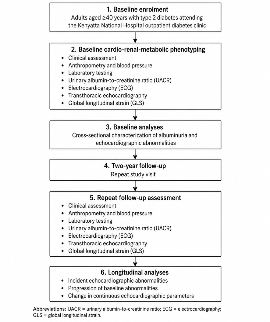
The MOYO-FIGO study is a prospective observational cohort study designed to evaluate whether albuminuria predicts progression of CV structural and functional abnormalities among adults with T2DM in Kenya (Figure 1). This manuscript describes the protocol for the longitudinal follow-up of an established cohort in which baseline CKM phenotyping has already been completed and two-year follow-up assessment is planned.
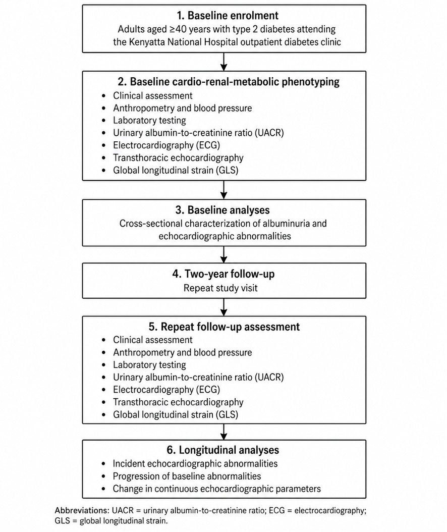
The study includes completed baseline CKM phenotyping and a planned two-year follow-up assessment. At baseline, participants underwent standardized clinical assessment, laboratory testing, urinary albumin-to-creatinine ratio measurement, and comprehensive transthoracic echocardiography, including global longitudinal strain assessment. Baseline assessment was conducted between August 2025 and February 2026. Participants will be invited for repeat clinical, laboratory, urinary, and echocardiographic assessment two years after baseline.
The primary longitudinal objective is to determine whether baseline albuminuria is associated with two-year change in global longitudinal strain. Secondary longitudinal analyses will evaluate incident and progressive cardiac structural and functional abnormalities over two years.
Study setting
The study is being conducted at the outpatient diabetes clinic of Kenyatta National Hospital in Nairobi, Kenya. Kenyatta National Hospital is the largest national referral and teaching hospital in Kenya and provides specialist outpatient diabetes care as well as cardiology and echocardiography services. This setting is therefore appropriate for evaluating CKM risk among adults with T2DM attending tertiary outpatient care, while recognizing that findings from this tertiary referral setting may not be fully generalizable to adults with T2DM managed in rural, primary-care, or community-based settings.
Study population
The study population consists of adults aged 40 years or older with documented T2DM attending routine follow-up at the Kenyatta National Hospital outpatient diabetes clinic. The age restriction was selected to enrich the cohort for participants in whom albuminuria and cardiac structural or functional abnormalities are more likely to be clinically detectable, reflecting longer cumulative exposure to hyperglycaemia and other cardiometabolic risk factors.
Eligibility criteria
Inclusion criteria
Participants were eligible for enrolment if they met all of the following criteria:
-
aged 40 years or older;
-
documented diagnosis of T2DM;
-
attending the Kenyatta National Hospital outpatient diabetes clinic;
-
able to understand the study procedures and provide written informed consent;
-
willing to undergo clinical assessment, blood sampling, urine collection, electrocardiography, and transthoracic echocardiography.
Exclusion criteria
Participants were excluded if they met any of the following criteria:
-
unable to provide informed consent because of cognitive impairment or other limiting condition;
-
clinically unstable or too unwell to complete study procedures;
-
known congenital heart disease or significant primary valvular heart disease;
-
prior history of myocardial infarction;
-
previous cardiac surgery, pacemaker, or implantable defibrillator;
-
end-stage kidney disease or current dialysis;
-
current pregnancy or less than six months postpartum;
-
current infection or fever, including suspected urinary tract infection;
-
current use of drugs likely to substantially affect albuminuria assessment, such as chemotherapy or high-dose non-steroidal anti-inflammatory drugs;
-
prior enrolment in the study.
Recruitment and cohort enrolment
Participants were recruited from adults with T2DM attending the Kenyatta National Hospital outpatient diabetes clinic during routine clinic visits. On recruitment days, a list of potentially eligible patients was obtained from clinic records using hospital numbers. This list served as the clinic-day sampling frame.
Simple random sampling without replacement was then performed using R statistical software to ensure that each eligible patient had an equal probability of selection and that no participant was selected more than once. Selected patients were approached sequentially using their hospital numbers, screened for eligibility, and invited to participate. Written informed consent was obtained before any study-specific procedures were conducted.
Participants who completed baseline clinical, laboratory, urinary albumin-to-creatinine ratio testing, electrocardiography, and echocardiographic assessment constitute the baseline cohort and will be invited for repeat assessment two years after their baseline visit.
Baseline clinical assessment
At baseline, all enrolled participants underwent standardized same-day clinical, laboratory, urinary, electrocardiographic, and echocardiographic assessment. Trained study staff collected socio-demographic data, medical history, diabetes duration, medication history, lifestyle factors, and relevant clinical information using a standardized electronic questionnaire.
Anthropometric measurements included weight, height, body mass index, waist circumference, and waist-to-hip ratio. Body mass index was calculated as weight in kilograms divided by height in metres squared. Blood pressure was measured using standardized procedures after the participant had rested in a seated position, consistent with recommendations for accurate blood pressure measurement in humans.13 Where repeated measurements were available, the average blood pressure value was used for analysis. Hypertension was defined based on prior clinical diagnosis, current use of antihypertensive treatment, or measured blood pressure meeting prespecified study thresholds.
Laboratory assessment
Venous blood samples were collected during the baseline study visit on the same day as urine collection and echocardiographic assessment. Laboratory tests included glycated haemoglobin, serum creatinine, lipid profile, and N-terminal pro-B-type natriuretic peptide.
At baseline, estimated glomerular filtration rate (eGFR) was calculated using the Chronic Kidney Disease Epidemiology Collaboration equation and categorized according to Kidney Disease: Improving Global Outcomes glomerular filtration rate categories.7 Lipid profile measurements included total cholesterol, low-density lipoprotein cholesterol, high-density lipoprotein cholesterol, and triglycerides.
At the two-year follow-up visit, clinical and laboratory measurements will be repeated using comparable procedures to evaluate change over time and to support longitudinal analysis of CKM risk.
Albuminuria measurement
Albuminuria was assessed using the UACR measured from a random spot urine sample collected during the baseline study visit. A random urine sample was selected because it is practical and feasible in the outpatient clinic setting. Participants were asked about intense physical activity in the preceding 24 hours and were instructed on appropriate urine sample collection procedures.
Urine samples were collected in sterile containers, sealed, stored in a cool environment, and transported to the laboratory within two hours of collection. Urine albumin and urine creatinine were measured from the same random spot urine sample using the laboratory’s standard chemistry platform. Urine albumin concentration was measured using an immunoturbidimetric immunoassay, while urine creatinine concentration was measured using a colorimetric method.
The UACR was calculated as urine albumin concentration divided by urine creatinine concentration and expressed as mg/g.
Echocardiographic assessment
All participants underwent comprehensive transthoracic echocardiography at baseline using the Vivid IQ Ultra Edition ultrasound system. Echocardiographic examinations were performed according to standardized acquisition protocols and included assessment of cardiac structure, systolic function, diastolic function, right ventricular systolic function, and myocardial deformation.
LV structural assessment included measurement of LV dimensions, LV mass, and LV mass index.
LVEF was measured using the modified Simpson’s biplane method from apical four-chamber and two-chamber views.14 LV diastolic function (LVDD) was assessed using mitral inflow velocities, tissue Doppler early diastolic mitral annular velocity, average E/e′ ratio, left atrial volume index, and peak tricuspid regurgitation velocity. LVDD will be classified using the 2016 American Society of Echocardiography and European Association of Cardiovascular Imaging recommendations.15
GLS was assessed using two-dimensional speckle-tracking echocardiography from standard apical views. Strain analysis was performed using the integrated Automated Functional Imaging left ventricular package on the Vivid IQ Ultra Edition system. GLS was calculated as the average peak systolic longitudinal strain across LV segments and expressed as a negative percentage, consistent with standard terminology for two-dimensional speckle-tracking echocardiography.16
Right ventricular systolic function was assessed using tricuspid annular plane systolic excursion from the right ventricular-focused apical four-chamber view.17
At the two-year follow-up visit, transthoracic echocardiography will be repeated using the same standardized protocol to evaluate incident abnormalities, progression of baseline abnormalities, and change in continuous echocardiographic parameters.
Exposure definition
The main exposure is baseline albuminuria, measured using the UACR. Albuminuria will be analysed categorically using Kidney Disease: Improving Global Outcomes albuminuria categories: A1, normal to mildly increased albuminuria, defined as urinary albumin-to-creatinine ratio <30 mg/g; A2, moderately increased albuminuria, defined as urinary albumin-to-creatinine ratio 30–300 mg/g; and A3, severely increased albuminuria, defined as urinary albumin-to-creatinine ratio >300 mg/g.7 UACR will also be analysed as a continuous variable, with logarithmic transformation considered if its distribution is markedly skewed.
For selected analyses, albuminuria may also be dichotomised as absent versus present, using urinary albumin-to-creatinine ratio <30 mg/g and ≥30 mg/g, respectively.7 This binary definition will be used only for secondary or sensitivity analyses, while the primary exposure definition will retain the three-level A1–A3 categorisation.
Outcome measures
Primary outcome
The primary outcome is two-year change in GLS, defined as the difference between global longitudinal strain measured at baseline and global longitudinal strain measured at the two-year follow-up visit. GLS will be assessed using two-dimensional speckle-tracking echocardiography and expressed as a negative percentage, consistent with standard terminology for myocardial deformation imaging. A less negative GLS value indicates worse left ventricular systolic deformation. For categorical analyses, abnormal GLS will be defined a priori as a value less negative than −16%.16
Secondary outcomes
Secondary outcomes will include incident or progressive abnormalities in the following echocardiographic domains:
-
left ventricular hypertrophy;
-
left atrial enlargement;
-
reduced left ventricular ejection fraction;
-
left ventricular diastolic dysfunction;
-
right ventricular systolic dysfunction;
-
any predefined echocardiographic abnormality.
LV hypertrophy (LVH) was defined using sex-specific LV mass index thresholds: >115 g/m² in men and >95 g/m² in women.14 Left atrial volume was measured and indexed to body surface area, with left atrial enlargement defined as left atrial volume index >34 mL/m².14 Reduced LVEF will be defined as LVEF <50%, measured using the modified Simpson’s biplane method.14
LV diastolic dysfunction will be assessed using the 2016 American Society of Echocardiography and European Association of Cardiovascular Imaging recommendations. The four variables used in the diagnostic algorithm will include septal e′ velocity <7 cm/s or lateral e′ velocity <10 cm/s, average E/e′ ratio >14, left atrial volume index >34 mL/m², and peak tricuspid regurgitation velocity >2.8 m/s.15
Right ventricular systolic dysfunction will be defined using tricuspid annular plane systolic excursion <1.7 cm, measured from the right ventricular-focused apical four-chamber view.17
Exploratory outcomes
Exploratory outcomes will include associations between stored serum proteomic or metabolomic profiles and albuminuria, global longitudinal strain change, and selected echocardiographic changes over follow-up. These analyses will be conducted only if permitted by ethics approval, funding availability, and participant consent provisions.
Follow-up assessment
Participants enrolled at baseline will be invited for a repeat study visit two years after their baseline assessment. The follow-up visit will repeat the core baseline procedures, including clinical assessment, blood pressure measurement, anthropometry, laboratory testing, UACR measurement, electrocardiography, and transthoracic echocardiography.
The repeat echocardiographic assessment will use the same standardized protocol as the baseline visit, including chamber quantification, LVEF, LVDD assessment, global longitudinal strain, and right ventricular systolic function assessment. This repeated assessment will allow evaluation of incident cardiac abnormalities, progression of baseline abnormalities, and change in continuous echocardiographic parameters over time.
Participants who cannot attend the follow-up visit will be contacted using available study contact information. Reasons for non-attendance, withdrawal, loss to follow-up, or death, where known, will be documented to support interpretation of longitudinal analyses.
Retention strategies
Several strategies will be used to maximize retention at the two-year follow-up visit. At baseline, participant contact information, including telephone numbers and alternative contacts where available, was collected to support future follow-up. Before the scheduled follow-up window, participants will be contacted by telephone and reminded about the repeat study visit. Where possible, follow-up visits will be aligned with routine diabetes clinic appointments to reduce additional travel and time burden. Participants who miss scheduled follow-up appointments will be contacted again and offered an alternative appointment date. Reasons for non-attendance, withdrawal, loss to follow-up, or death, where known, will be documented. Baseline characteristics of participants retained in follow-up will be compared with those lost to follow-up to assess potential attrition bias.
Biospecimen storage and exploratory omics analyses
During the baseline visit, serum samples were collected as part of the study laboratory assessment. Subject to availability of funding, additional ethics approval where required, and participant consent provisions, stored serum samples may be used for exploratory proteomic and metabolomic analyses. These analyses will be exploratory and hypothesis-generating, with the aim of identifying circulating molecular signatures associated with albuminuria, cardiac structural abnormalities, myocardial dysfunction, and progression of CKM abnormalities over follow-up.
Data management and quality assurance
Study data were collected directly into a REDCap electronic database using tablet devices. REDCap is a secure, web-based software platform designed to support electronic data capture for research studies.18 Direct electronic data entry was used to reduce transcription errors, support real-time data validation, and allow routine data quality monitoring.
The database included structured electronic case report forms, programmed field validations, range checks, skip patterns, and user-specific access controls. REDCap also maintains an audit trail of data entry and modification, allowing changes to be tracked by user and time.18 Access to the database was restricted to authorized study personnel using individual login credentials. Tablets used for data collection were password protected and stored securely after each data collection session.
Data quality assurance procedures were conducted throughout baseline data collection. Automated and interactive quality control reports were generated using R software to identify missing values, inconsistent entries, outliers, and other potential data issues. R is a statistical computing environment used for data management, statistical analysis, and graphics.19 Data queries were reviewed by the study team and corrected using source information where appropriate. Study progress reports were also generated to monitor recruitment, completeness of study procedures, and operational challenges.
For the imaging component, echocardiographic acquisition followed a standardized study protocol. Examinations were performed by an experienced cardiac sonographer, with interpretation and validation by an expert in echocardiography. The same echocardiographic definitions and measurement protocols will be used at baseline and follow-up to improve consistency in longitudinal assessment.
Sample size considerations
The baseline sample size was determined for the initial descriptive and analytic objectives of the MOYO-FIGO cohort. For the descriptive objective estimating albuminuria prevalence, the single population proportion formula was used, assuming an expected microalbuminuria prevalence of 37.11% among diabetes patients in Africa, based on a systematic review and meta-analysis.11 With a 95% confidence level (CI), 8% precision, and 10% allowance for non-response or incomplete data, the adjusted sample size required for this objective was 155 participants. For the descriptive objective estimating echocardiographic abnormalities, the same single population proportion formula was applied using published estimates of echocardiographic abnormalities among adults with T2DM. These included left ventricular remodeling, left ventricular diastolic dysfunction, impaired global longitudinal strain, and left atrial enlargement from a study in Cameroon,20 and LVDD from a systematic review and meta-analysis of adults with T2DM.21 The largest required sample size was 165 participants, based on an expected prevalence of LVDD of 51.6%.20
For the analytic objective assessing the association between albuminuria and echocardiographic abnormalities, published albuminuria-stratified data were reviewed. Jørgensen et al. reported associations between albuminuria severity and cardiac mechanics in T2DM, but many of the relevant measures were reported as continuous echocardiographic parameters.12 Therefore, categorical albuminuria-stratified LVDD data from a sub-Saharan African T2DM population were used for the formal analytic sample size estimation.22 Using a chi-square test with Cramer’s V = 0.256, α = 0.05, 80% power, and 2 degrees of freedom, the minimum analytic sample size was 147 participants. After allowing for 10% non-response or incomplete data, the adjusted analytic sample size was 162 participants. This calculation was performed using the pwr package in R.19
The final minimum baseline sample size was therefore set at 165 participants, corresponding to the largest requirement across the descriptive and analytic objectives. For the planned longitudinal analysis, all participants who completed baseline assessment will be invited for the two-year follow-up visit. A separate longitudinal sample size calculation was not performed because reliable local estimates of two-year progression rates in echocardiographic abnormalities across albuminuria categories were not available. Longitudinal analyses will therefore use all available follow-up data and and will be interpreted with emphasis on effect estimates with 95% CIs rather than statistical significance alone. Follow-up completeness will be reported, and baseline characteristics of participants retained versus lost to follow-up will be compared to assess potential attrition bias. If follow-up numbers are smaller than anticipated, analyses of less frequent secondary outcomes will be considered exploratory.
Statistical analysis plan
Analyses will be conducted in two stages: baseline analyses and longitudinal analyses. Baseline analyses will characterize the established cohort at the time of first assessment and will be reported separately from the longitudinal follow-up analysis. Longitudinal analyses will evaluate whether baseline albuminuria is associated with two-year change in global longitudinal strain, the primary longitudinal outcome, and with selected secondary echocardiographic outcomes at follow-up.
Baseline participant characteristics will be summarized overall and by albuminuria category. Categorical variables will be described using frequencies and percentages. Continuous variables will be summarized using means and standard deviations when approximately normally distributed, and medians with interquartile ranges when skewed. Baseline comparisons across albuminuria categories will use chi-square or Fisher’s exact tests for categorical variables and analysis of variance or Kruskal–Wallis tests for continuous variables, as appropriate.
The baseline prevalence of each predefined echocardiographic abnormality will be estimated as the proportion of participants meeting the operational definition for that abnormality. These abnormalities will include LVH, left atrial enlargement, reduced LVEF, abnormal global longitudinal strain, LVDD, right ventricular systolic dysfunction, and any abnormal echocardiographic finding. Prevalence estimates will be reported with 95% CIs.
For baseline cross-sectional analyses, logistic regression models will be used to estimate the association between albuminuria category and each binary echocardiographic abnormality. Normal to mildly increased albuminuria, defined as UACR of less than 30 mg/g will serve as the reference category. Crude odds ratios and adjusted odds ratios will be reported with 95% CIs. Multivariable models will adjust for prespecified clinically relevant covariates, including age, sex, hypertension, duration of diabetes, body mass index, and glycated haemoglobin. Additional adjustment for eGFR may be considered in sensitivity analyses, recognizing that kidney function may lie on the causal pathway between albuminuria and cardiovascular outcomes.
For the primary longitudinal analysis, the outcome will be two-year change in global longitudinal strain. Baseline albuminuria will be the main exposure and will be analysed using A1, A2, and A3 categories, with A1 as the reference category. Additional analyses will assess urinary albumin-to-creatinine ratio as a continuous exposure, with logarithmic transformation considered if the distribution is markedly skewed. Linear regression will be used to estimate the association between baseline albuminuria and change in global longitudinal strain, adjusting for prespecified covariates including age, sex, hypertension, duration of diabetes, body mass index, glycated haemoglobin, and baseline global longitudinal strain. If repeated measures are modelled jointly, linear mixed-effects models may be used. Because albuminuria at baseline was assessed using a single random spot UACR, sensitivity analyses will consider repeat UACR measured at follow-up where available. These analyses may classify participants according to persistent albuminuria, regression of albuminuria, progression of albuminuria category, or change in UACR over time. These analyses will be considered exploratory and will be interpreted cautiously because follow-up UACR may reflect both baseline risk and interval changes in kidney status.
Secondary longitudinal analyses will evaluate incident or progressive echocardiographic abnormalities over two years. Logistic regression will be used for binary incident outcomes, while linear regression or linear mixed-effects models will be used for change in continuous echocardiographic parameters. Participants lost to follow-up will be compared with those retained in the cohort using baseline characteristics to assess potential attrition bias.
Missing data will be assessed for extent, and likely pattern. Available-case analysis will be used for primary analyses when missingness is limited. If missing data are substantial, sensitivity analyses will be considered to assess the robustness of findings, , including multiple imputation where appropriate and where assumptions are reasonable. Participants lost to follow-up will be compared with those retained in the cohort using baseline characteristics to evaluate potential attrition bias.
Model assumptions will be assessed before interpretation of regression models. For linear regression models, residual distribution, linearity, influential observations, and homoscedasticity will be examined. For logistic regression models, sparse outcome categories, influential observations, and model convergence will be assessed. Continuous covariates will be checked for functional form, and transformation or flexible modelling will be considered where appropriate.
Covariates for multivariable models will be selected a priori based on clinical relevance and plausible confounding of the relationship between albuminuria and cardiac structural or functional outcomes. Prespecified covariates will include age, sex, hypertension, duration of diabetes, body mass index (BMI), glycated haemoglobin (HbA1c), and baseline GLS for the primary longitudinal analysis. eGFR will be considered in sensitivity analyses because kidney function may be related to both albuminuria and cardiovascular outcomes and may also lie on the causal pathway.
Given the number of secondary and exploratory outcomes, these analyses will be interpreted with attention to multiplicity and consistency of effect estimates rather than isolated p-values. The primary longitudinal outcome will be two-year change in GLS. Secondary and exploratory analyses will be considered hypothesis-generating, and findings will be reported with effect estimates and 95% CI.
All statistical analyses will be conducted using R software.19 Statistical tests will be two-sided, and p-values less than 0.05 will be considered statistically significant.
DISCUSSION
The MOYO-FIGO study is designed to generate longitudinal follow-up of an established cohort with completed baseline CKM phenotyping. The study will generate longitudinal evidence on whether albuminuria is associated with two-year change in GLS among adults with T2DM in Kenya, while also evaluating secondary structural and functional echocardiographic outcomes. By combining UACR testing with comprehensive echocardiography, including global longitudinal strain, MOYO-FIGO is designed to evaluate whether albuminuria can serve as a practical marker for identifying adults with T2DM who are at higher risk of progressive cardiac involvement.
A major contribution of this cohort is its focus on CKM risk in an African tertiary outpatient diabetes population. Albuminuria is an established marker of kidney involvement and is also associated with CV events, heart failure, and mortality.9,10 Previous studies have reported associations between albuminuria and cardiac mechanics in T2DM, but longitudinal African data integrating albuminuria with broad echocardiographic phenotyping remain limited.12 MOYO-FIGO is therefore positioned to address an important evidence gap by evaluating whether baseline albuminuria predicts incident or progressive cardiac abnormalities over two years. The study also has relevance for global health economics and policy. Echocardiography and strain imaging require specialized equipment and trained personnel and may not be routinely available for all adults with T2DM in resource-limited settings. If albuminuria is shown to identify patients at higher risk of cardiac structural or functional progression, UACR testing could support more targeted referral for echocardiography. MOYO-FIGO will not directly evaluate cost-effectiveness, but the findings may provide empirical inputs for future economic evaluations of albuminuria-guided cardiovascular screening strategies in diabetes care.
The inclusion of GLS is important because early myocardial dysfunction in T2DM may not be captured by LVEF alone. Conventional echocardiographic assessment often relies on LVEF, but ejection fraction may remain preserved during early myocardial dysfunction. GLS provides a more sensitive assessment of myocardial deformation and may detect subclinical left ventricular systolic dysfunction before overt reduction in ejection fraction becomes apparent6. In a setting where echocardiography resources are limited, identifying clinical or laboratory markers that can help target patients for more detailed CV assessment may be valuable. MOYO-FIGO will not establish whether albuminuria-based screening is cost-effective, but it may provide evidence to support early identification of patients who warrant closer cardiovascular assessment and integrated CKM risk management.
STRENGTHS AND LIMITATIONS
The study has several strengths. First, it uses a prospective observational cohort design with completed baseline CKM phenotyping and planned two-year follow-up. Second, the baseline assessment integrates clinical data, laboratory testing, UACR measurement, electrocardiography, and echocardiography within the same study visit. Third, the echocardiographic protocol includes chamber quantification, systolic function, diastolic function assessment, right ventricular systolic function, and GLS. Fourth, data were captured electronically using REDCap, with programmed validation checks and routine quality assurance procedures.
The study also has limitations. It is single-centre cohort based at Kenyatta National Hospital, a national tertiary referral and teaching hospital. Participants attending this clinic may differ from adults with T2DM managed in rural, primary-care, county-hospital, or community-based settings. They may have longer disease duration, greater comorbidity burden, more advanced complications, or better access to specialist investigations than patients outside a tertiary referral system. Therefore, the findings may not be directly generalizable to all adults with T2DM in Kenya.
Albuminuria was measured at baseline using a single random spot UACR. Although this approach is practical in an outpatient clinic setting, persistent albuminuria cannot be confirmed without repeat testing.
This may lead to exposure misclassification because urinary albumin excretion can vary with hydration status, exercise, infection, glycaemic control, blood pressure, and intercurrent illness. Repeat UACR measurement at follow-up will allow exploratory analyses of persistent, regressing, or progressing albuminuria categories where data are available.
The longitudinal analyses may also be affected by attrition over the two-year follow-up period. Participants who return for repeat assessment may differ systematically from those who are lost to follow-up, potentially introducing attrition bias. To address this, the study will use participant contact and reminder procedures, document reasons for non-attendance where known, and compare baseline characteristics of participants retained versus lost to follow-up. The sample size may limit power for less frequent secondary outcomes and subgroup analyses, and these analyses will be interpreted cautiously.
Finally, because MOYO-FIGO is observational, it can evaluate associations and temporal patterns but cannot establish causality.
CONCLUSIONS
MOYO-FIGO is a prospective observational cohort study describing the longitudinal follow-up of an established cohort with completed baseline CKM phenotyping and planned two-year follow-up. The study is designed to evaluate whether albuminuria is associated with two-year change in global longitudinal strain and secondary echocardiographic outcomes among adults with T2DM attending a tertiary outpatient clinic in Kenya. Findings from this cohort may inform future strategies for risk stratification, targeted echocardiographic assessment, integrated CKM care , and future economic evaluation of albuminuria-guided cardiovascular screening strategies in resource-limited diabetes settings.
Acknowledgements
The authors acknowledge the participants who agreed to take part in the MOYO-FIGO study. We also thank the staff of the Kenyatta National Hospital outpatient diabetes clinic, cardiology/echocardiography unit, and laboratory partners for their support during baseline data collection.
Ethics and dissemination
Ethical approval for the baseline MOYO-FIGO assessment was obtained from the Kenyatta National Hospital–University of Nairobi Ethics Review Committee, approval number P713/09/2024. Written informed consent was obtained from all participants before enrolment. The study was explained in the participant’s preferred language, English or Kiswahili, including the study purpose, procedures, potential risks and benefits, voluntary nature of participation, and the right to decline or withdraw without affecting clinical care.
The risks associated with baseline study procedures were minimal and mainly related to minor discomfort during blood sampling, urine collection, and echocardiography. Participant confidentiality was maintained using coded identifiers, restricted access to study data, password-protected electronic systems, and secure data storage. Clinically important incidental findings identified during study procedures were communicated to the treating clinical team for appropriate care within Kenyatta National Hospital.
The planned two-year follow-up assessment, including repeat clinical, laboratory, UACR, and echocardiographic evaluation, will be conducted subject to ethics approval or amendment and appropriate participant consent provisions. Any future exploratory proteomic or metabolomic analyses using stored serum samples will also be conducted only if permitted by ethics approval, funding availability, and participant consent provisions.
Findings from the MOYO-FIGO study will be disseminated through peer-reviewed publications, scientific conferences, and presentations to relevant clinical and academic stakeholders. Study outputs will also be shared with the University of Nairobi Department of Clinical Medicine and Therapeutics, relevant clinical departments at Kenyatta National Hospital, and the Kenyatta National Hospital Research Department. No individual-level identifiable participant data will be disclosed in any dissemination output.
Data availability statement
The dataset generated from the MOYO-FIGO study will not be publicly released with individual-level identifiers. De-identified data may be made available by the corresponding author upon reasonable request and subject to institutional approval, ethics requirements, and applicable data-sharing agreements.
Funding statement
The baseline MOYO-FIGO assessment did not receive dedicated external funding. Additional funding will be sought to support the planned two-year follow-up assessment and exploratory proteomic and metabolomic analyses.
Disclosure of interest
The authors completed the ICMJE Disclosure of Interest Form and declare no relevant competing interests.
Authors’ contributions
BMA conceived the study, developed the protocol, coordinated study implementation, and drafted the manuscript. ENO, CFO, NABN, and CTL provided scientific supervision and clinical guidance. DM, AB, POO, and AM contributed to manuscript review. All authors reviewed and approved the final manuscript.
Disclosure of interest
The authors declare no competing interests.
Correspondence to:
Boni Maxime Ale, MD, MSc, MPH
Cardiovascular & Metabolic Research Center,
Holo Global Health Research Institute
Nairobi, Kenya
Email: bale@holohc.org